It is the goal of the Electron Microscopy, Histopathology and Tissue Bank Core to provide the research community with the technical services necessary for growth and development in the world of investigative research. The services that will be provided by the Core are essential to an institution upholding a status of high recognition nationally and internationally. At a time when new discoveries generated by basic biomedical research are applied to pathology practice, accessibility to the most modern scientific methods becomes imperative.
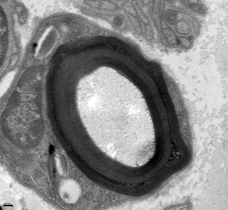
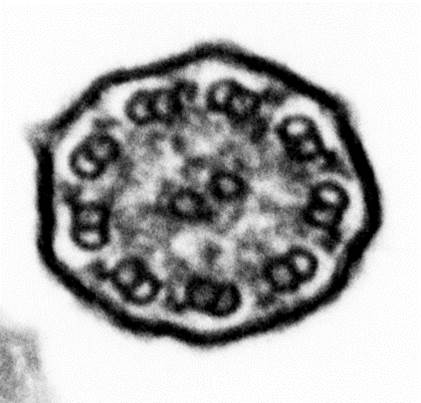
Electron Microscopy
Histopathology
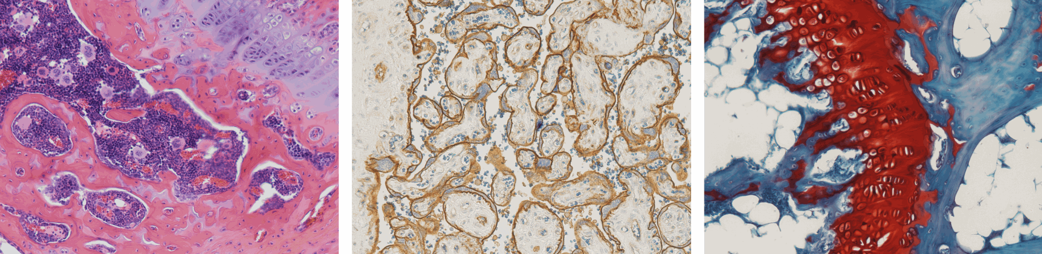
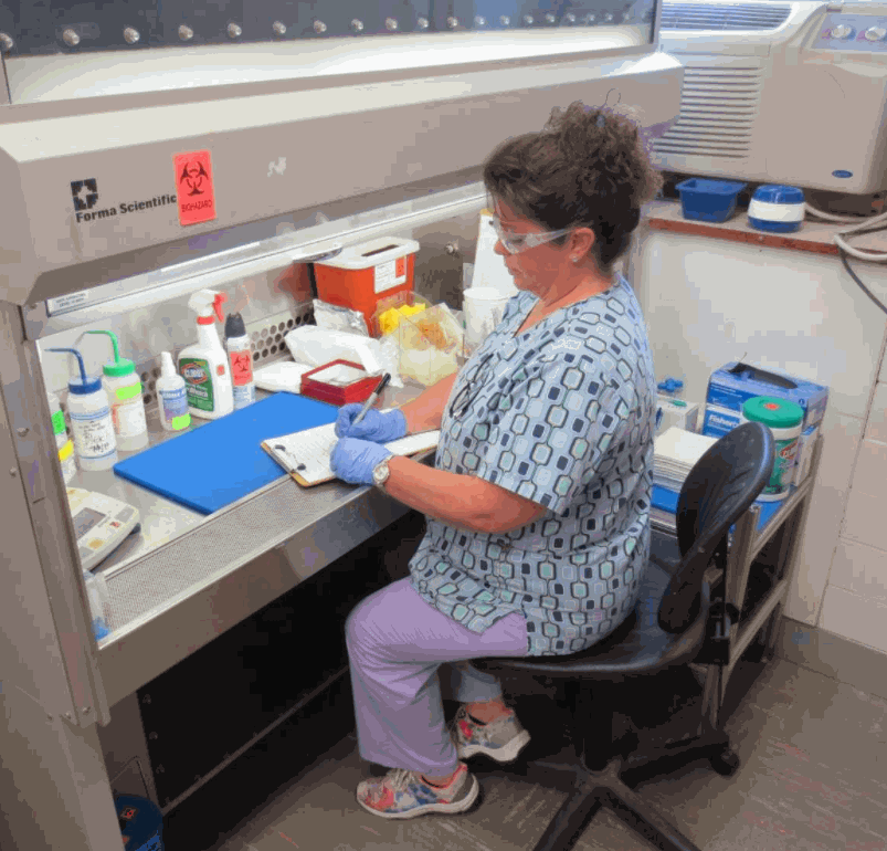
The Pathology Research Histology Laboratory assists the research community by providing access to high quality, timely and cost-effective histologic services. Available routine histologic procedures include gross processing of tissue and cellular specimens, paraffin embedding, sectioning, and staining (routine histologic and immunohistochemical stains). Advanced services, including development of specialized histologic stains and tissue microarray construction, are available in consultation with the laboratory director.
Tissue Bank and Brain Bank
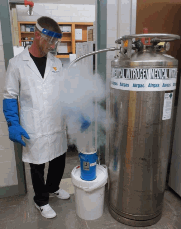
Tissues are obtained fresh, frozen in liquid nitrogen, embedded in the cryopreservative O.C.T., formalin fixed paraffin embedded, and/or using specific collection media provided by the researcher
Peng Han, M.D., Ph.D
Assistant Professor, Pathology, Anatomy and Laboratory Medicine
Adjunct Faculty, Department of Neuroscience
Principal Investigator, Tissue Bank and Brain Bank
Laboratory Director, Research Histology
304-293-0645
peng.han@hsc.wvu.edu
David Howell, M.D., Ph.D
Assistant Professor, Pathology, Anatomy and Laboratory Medicine
Electron Microscopy Laboratory Director
304-293-1628
dhowell@hsc.wvu.edu
Monday - Friday
6:00 AM - 4:00 PM
https://medicine.hsc.wvu.edu/pathology/
https://medicine.hsc.wvu.edu/pathology/research/pathology-research-laboratory/
WVU HSC Core Facilities
| Name | Role | Phone | Location | |
|---|---|---|---|---|
| David Howell |
Director, Electron Microscopy
|
304-293-1628
|
dhowell@hsc.wvu.edu
|
2115 HSC N
|
| David Huffman |
Supervisor, Electron Microscopy
|
304-293-6716
|
dhuffma7@wvumedicine.org
|
2114 HSC N
|
| Jacqueline Karakiozis |
Supervisor, Tissue Bank and Brain Bank & Pathology Research Histology
|
304-293-0287
|
jkarakiozis@hsc.wvu.edu
|
2126 HSC N
|
| Phil Han |
Director, Tissue Bank and Brain Bank & Pathology Research Histology
|
304-293-0645
|
peng.han@hsc.wvu.edu
|
| Service list |
| ► Tissue Bank (8) | |||
| Name | Description | Price | |
|---|---|---|---|
| Additional Abstracting/Technical service | Inquire | ||
| Database Query | Inquire | ||
| FFPE Block | Inquire | ||
| Fresh Tissue - Malignant | Inquire | ||
| Fresh Tissue - Non-Malignant | Inquire | ||
| Frozen Tissue (malignant and non-malignant) | Inquire | ||
| Matching blood, plasma, or serum | Inquire | ||
| Quality Assurance Determination (Tumor vs Normal) | Inquire | ||